Ocular and periocular diagnoses: Difference between revisions
m (Rossdonaldson1 moved page Eye algorithms (main) to Ocular and periocular diagnoses) |
No edit summary |
||
| Line 5: | Line 5: | ||
[[File:eyelid glands.png|thumb]] | [[File:eyelid glands.png|thumb]] | ||
== | ==Clinical Features== | ||
{{Eye images}} | |||
{{Conjunctivitis images}} | |||
==Differential Diagnosis== | |||
[[File:Red_Eye_-_Advanced.png|thumbnail|Algorithm for the evaluation of red/painful eye]] | [[File:Red_Eye_-_Advanced.png|thumbnail|Algorithm for the evaluation of red/painful eye]] | ||
{{Eye algorithms}} | {{Eye algorithms}} | ||
==[[ | ==Evaluation== | ||
*[[Eye exam]] | |||
==Management== | |||
*Diagnosis dependent | |||
==Disposition== | |||
*Diagnosis dependent | |||
==See Also== | ==See Also== | ||
*[[ | *[[Diagnoses by Body Part (Main)]] | ||
==External Links== | ==External Links== | ||
Revision as of 20:58, 23 October 2024
Background


Clinical Features
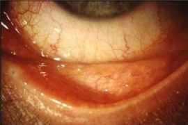
Eye images
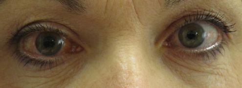
Acute angle closure glaucoma (right)
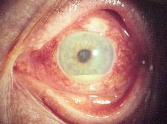
Anterior uveitis with hypopyon
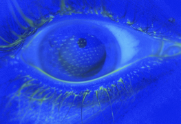
Airbag corneal abrasion
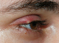
External stye
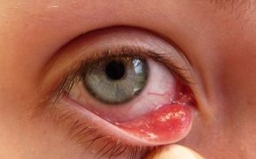
Internal stye


Conjunctivitis Images
Acute allergic conjunctivitis
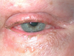
Chronic allergic conjunctivitis
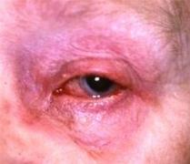
Contact blepharoconjunctivitis


Differential Diagnosis

Eye Algorithms
- Red eye
- Periorbital swelling
- Acute vision loss (noninflamed)
- Acute onset flashers and floaters
- Painful eyes with normal exam
- Neonatal eye problems
Evaluation
Management
- Diagnosis dependent
Disposition
- Diagnosis dependent
See Also
External Links
- Summary review: http://www.emdocs.net/approach-to-the-red-eye/
- Central vs Peripheral Vision loss algorithms: http://www.emdocs.net/central-vs-peripheral-vision-loss/